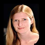
Professor of Biomedical Engineering and Radiology
Zuckerman Mind Brain Behavior Institute at Columbia University
Using Light to Image at the Speed of Life
Abstract
Light is uniquely capable of probing and changing the electron and vibrational energy levels of molecules. As a result, light can be used to spectrally discern a tissue’s chemical composition, excite activity in a neuron and change the shape of the cornea in laser eye surgery. As lasers, light-sources, detectors and cameras continue to improve, the opportunities to leverage the power of light for biomedical discovery, diagnosis and treatment are continually expanding.
An area of enormous growth in the past decade has been the development of fluorescent protein-based reporters of cellular activity and optogenetics, which combined have unlocked our ability to observe and manipulate cell signaling across scales. The result is a rich and expanding toolkit for studying how intact, living organs, organoids and organisms function, and how they are affected by disease, which in turn is fueling revolutions in neuroscience, drug discovery, personalized medicine. However, a major bottleneck for these techniques is imaging speed. We developed swept confocally aligned planar excitation (SCAPE) microscopy to address this need for speed, enabling 3D imaging in living tissues at over 100 volumes per second. SCAPE combines the high signal to noise and low photobleaching benefits of light sheet imaging with confocal descanning principles that enable high-speed scanning through a simple single, stationary objective lens. Through diverse collaborations, we have demonstrated SCAPE’s ability to image cellular activity in a wide range of living organisms including freely crawling Drosophila larvae, the whole brain of behaving adult Drosophila, zebrafish brain and heart, 3D culture systems and the awake mouse cortex. We are now developing a range of new SCAPE systems including two-photon, higher resolution and meso-scale versions, as well as platforms for high-throughput and high-content imaging of cleared and expanded tissues, and a miniaturized version of SCAPE for in-situ histopathology for clinical surgical guidance.
SCAPE is one example of our innovations that have continually sought to leverage the power of light to capture in-vivo dynamics. In parallel we have developed image analysis methods for spatiotemporal and hyperspectral unmixing enabling us to relate complex multidimensional observations to physiological phenomena and behavior. We are applying these tools in our own studies of real-time brain-wide neural activity, and to explore the relationship between blood flow and neural activity in the brain, seeking biomarkers and therapeutic targets for neurovascular dysfunction.
Biography
Dr Elizabeth Hillman is a Professor of Biomedical Engineering and Radiology, and a member of the Zuckerman Mind Brain Behavior Institute at Columbia University. Dr Hillman completed her undergraduate degree in Physics, and PhD in Medical Physics and Bioengineering at University College London. After a year at a Boston medical device start-up company, Dr Hillman completed postdoctoral training at the Martinos center for Biomedical Imaging at Massachusetts General Hospital / Harvard Medical School before becoming faculty at Columbia University in 2006. Prof Hillman has developed a range of novel approaches to in-vivo optical imaging and microscopy for both clinical and research applications. Her PhD work focused on time-resolved diffuse optical tomography of the breast and premature infant brain, while her innovations since have included Dynamic Contrast enhanced molecular imaging (DyCE, licensed to CRi Inc, now PerkinElmer), Laminar Optical Tomography for brain and skin imaging, hyperspectral two-photon microscopy, wide-field optical mapping (WFOM) of neuronal activity and hemodynamics and most recently high-speed 3D swept confocally aligned planar excitation (SCAPE) microscopy (now licensed to Leica Microsystems). Continuing technology development includes new variants of SCAPE including a miniaturized clinical version for ‘real-time’ intrasurgical in-situ histopathology. Dr Hillman also has an active research program applying her in-vivo imaging tools to studying neurovascular coupling and brain-wide resting state neural activity dynamics in the healthy, diseased and developing rodent brain. Dr Hillman has developed educational programs at Columbia University focusing on Disruptive Biomedical Design, Biomedical Imaging and Advanced Microscopy. She is a Fellow of the OSA, SPIE and AIMBE, and her awards include an NSF CAREER Award in 2010, the 2011 Optical Society of America Lomb Medal for contributions to optics at a young age, and the SPIE Biophotonics Technology Innovator award in 2018.
For more information or to schedule a meeting with the speaker, please contact Hanna Kim.
Hosted by Dr. Matt Brenner


