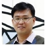
Woonggyu Jung, Ph. D.
Ulsan National Institute of Science and Technology, Korea
The Potential of Digital Histopathology Using Label-Free Optical Imaging Techniques
Abstract
The histological optical imaging is a gold standard method to observe the biological tissues, which follows routine process such as dissection, embedding, sectioning, staining, visualization and interpretation of specimens. This technique has a long history of development, and is used ubiquitously in pathology, despite being highly time and labour-intensive. Advanced optical imaging techniques developed over the last decade have enabled to provide high sensitivity, high resolution and non-invasive biological information. However, acquiring high throughput, large volume tissue anatomy remains a difficult challenge due to the effect of light scattering, which limits the penetration imaging depth and lateral resolution. Recently, various optical imaging methods have been introduced to create volumetric anatomy data of ex vivo tissues using physical tissue sectioning or optical clearing. Even though these new approaches present the distinguished volumetric anatomy in various scales, they are still not suitable for use in statistical studies with multiple tissues and organs. Here, we introduce novel label-free and multi-scale imaging modality based on serial optical coherence microscopy (OCM). OCM is a potential technique to build volumetric anatomy of mouse tissues or organs due to its simplicity, efficiency, robustness, and high-throughput capabilities. This presentation covers the latest work of large-scale brain and kidney imaging using OCM and its potential in bio-applications. Specifically, the talk will highlight other label-free optical imaging modalities including wide-field quantitative phase microscopy and optical projection tomography toward to multi-scale histopathology.
Biography
Woonggyu Jung received his Ph. D. in 2008 from the Department of Biomedical Engineering at the University of California, Irvine. From 2001 to 2008, he worked at the Beckman Laser Institute and Medical Clinic at UC Irvine. He also worked at the Beckman Institute for Advanced Science and Technology at the University of Illinois at Urbana-Champaign since January 2009. He has joined the faculty of UNIST in 2012, and currently works as an associate professor of Department of Biomedical Engineering. He is also co-founder and CTO of start-up company, Conecson which is focused on the futuristic business regarding to mobile-based medical devices. Dr. Jung has a strong research background in optical imaging technologies including optical coherence tomography (OCT), quantitative phase microscope (QPM), and miniaturized optical imaging probes. His research interest is to develop new optical technologies that address challenges in clinical medicine, basic biological research and neuroscience. In previous work, he developed a successful optical platform for in vivo translational research, and has published more than 60 peer-reviewed journal papers in the field of biophotoics.
For more information or to schedule a meeting with the speaker, please contact Xandra Dvornikova.
Sponsored by the Berns Family Laser and Microbeam Program
Hosted by: Dr. Zhongping Chen


