
Generating XACT images with a single-shot X-ray pulse in nanoseconds
By: Aili McConnon
Photo by: AIP Scilight
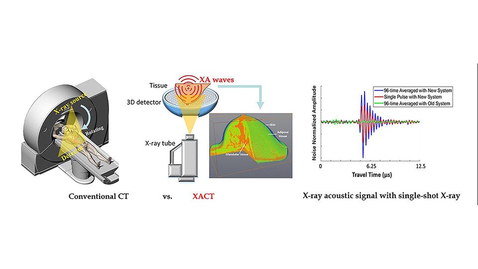
X-ray induced acoustic computed tomography can image the body at faster speeds and with smaller doses of radiation compared to traditional CT imaging
Siqi Wong, a graduate student in the lab of UCI Beckman Laser Institute & Medical Clinic’s Shawn Liangzhong, published a paper in Applied Physics Letters. The paper was chosen as an Editor’s Pick and highlighted by the American Institute of Physics.
X-ray imaging is widely deployed in scientific and medical realms, but the harm of radiation and slow imaging speed remain key limitations. Wang et al. developed a new type imaging modality called XACT (X-ray induced acoustic computed tomography) that is a promising alternative to traditional X-rays. XACT is similar to CT scans, but XACT can image the human body at a much faster speed and a much smaller radiation dose compared to traditional CT imaging.
“The innovation here is that we only need a single-shot X-ray pulse in nanoseconds to generate an XACT image, which typically requires around 1,000 pulses for averaging,” said author Siqi Wang.
The researchers used a 128-channel ring-shaped ultrasound array and a 150 kilovoltage peak X-ray source with a 50-nanosecond pulse width to achieve full tomographic imaging capability from a single pulse with a spatial resolution of 0.97 millimeters. The team plans to develop a portable 3D X-ray fluoroscopy, which could have a large impact in interventional radiology in clinical practice.
Click here to read the full article on AIP Scilight.

